|
To model the muscle composition, we used spaghetti and straws in different sizes. The spaghetti noodles represent the myofibrils. It is composed of long proteins which are in charge of sliding and contracting the muscles. A bundle of myofibrils forms a muscle fiber(black and thin straws), which are actual muscle cells with multiple nuclei, mitochondria, and a cellular membrane. The muscle fibers combine to form fascicle(colorful straws). Fascicles then combine to form the skeletal muscle represented by the white column in the picture. Every movement of our bodies requires the active engagement of our skeletal muscles. Learning muscle anatomy is important because we will have a more comprehensive understanding of the functions, location, and mechanism of muscles. 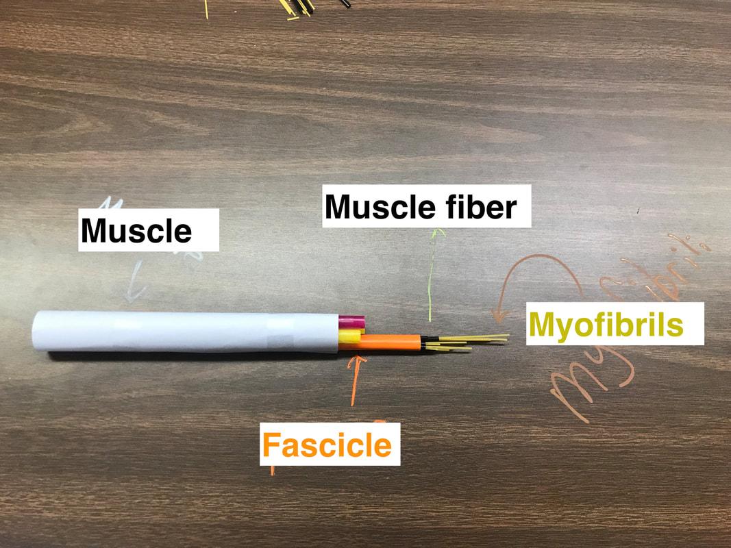
0 Comments
We removed the skin to expose the muscles, as shown in figure 3-1~3. The muscular system stabilizes joint, generates heat, and controls movement. We were able to identify pectoralis, obliques, latissimus dorsi, trapezius, bicep brachii, bicep femoris, and gastrocnemius(calf muscle). They each have different shapes. For example, the trapezius shapes like a trapezoid and allows scapula to rotate and lift. Biceps brachii, bicep femoris, and gastrocnemius are strong muscles on the limbs, they contract and help the movement of the limbs. Pectoralis are at the chest area and is responsible for shoulder movements. Left to right: 3-1 Pectoralis, oblique; 3-2 Biceps brachii; 3-3 latissimus, trapezius, biceps femoris, gastrocnemius Next, we cut the rat open along the midsagittal plane, from the neck to the urogenital opening(Figure 6-1~6). The thoracic and abdomen area include the cardiovascular, respiratory, digestive, and endocrine systems. The heart and lung(Figure 6-1) is part of the cardiovascular system, which transports nutrients, hormones, oxygen, and removes metabolic waste. The thymus gland(Figure 6-1) right above the heart and between the lungs is part of the immune system and helps produce white blood cells and fight diseases. The spleen(Figure 6-5) is also part of the lymphatic system, clearing our old red blood cells and other foreign bodies to help fight infection. The lungs(Figure 6-1) are the key structure in the respiratory system, it is the site of gas exchange, which takes in oxygen and expels CO2. The diaphragm(Figure 6-1) pulls down and lower the pressure inside the cavity, therefore, helps the gas “pump” into the lungs. The digestive system consists of the esophagus, stomach, pancreas, small and large intestine(Figure 6-2~4,6-6). They work together to break up and digest food and absorb nutrients. Moving on to the lower half of the abdomen cavity, step 7: the urogenital system, we collaborated with Sophia and Alexa’s group since they had a male rat. The urinary system removes liquid waste from the blood. We were able to locate the kidneys(Figure 7-1), urethra, and bladder(Figure 7-2). The reason why the bladder is so small that it’s hard to see is that the rat was not trained to hold urine for a long time. For the reproductive system, we were able to identify the vagina(Figure 7-2), uterine horns(Figure 7-1), and the ovaries(Figure 7-3), which produce the egg, protect and nourish the offspring. We visited Sophia and Alexa’s lab bench and they showed us the male reproductive system, the testes(Figure 7-5), scrotal sac, and the penis(Figure 7-4), which are responsible for producing and depositing sperms. Lastly, we removed the skin around the neck and exposed salivary glands(Figure 5-1) and lymph glands(Figure 5-2), as well as trachea(Figure 5-3) and larynx(Figure 5-4), the upper parts of the respiratory and digestive systems. Although the salivary glands(Figure 5-1) is a gland, it ]doesn’t belong to the endocrine system, it is actually one of the exocrine glands, secreting saliva that runs outside of the body. Its product, the saliva is also an important part of the digestive system, starting off the first step of digestion when the food enters the body. The lymph glands(Figure 5-2) belong to the immune system and are the major sites of white blood cell productions, protecting the body from infection. Hi! Welcome to the blog page where I keep notes and cool findings about anatomy and physiology! I was instantly attracted to this course because of anatomy -- I wanted to learn about all the body parts, their names, structures, and locations. Moreover, I am interested in the physiology -- the functions of each organs system, how the structures fit the functions, and how they work with each other to support the daily lives of the human beings, the most intricate machines on earth. So far, we started off by learning some terminologies of body orientations, body regions/cavity names, organ systems, and bone names. Though remembering all the big words can be tough, I had so much fun pointing at the manikins and being able to call out the “professional names” for each region :)
|
AuthorWelcome to my Anatomy & Physiology blog! Background Image credit:
https://www.mahipoweryoga.com/ ArchivesCategories |


 RSS Feed
