|
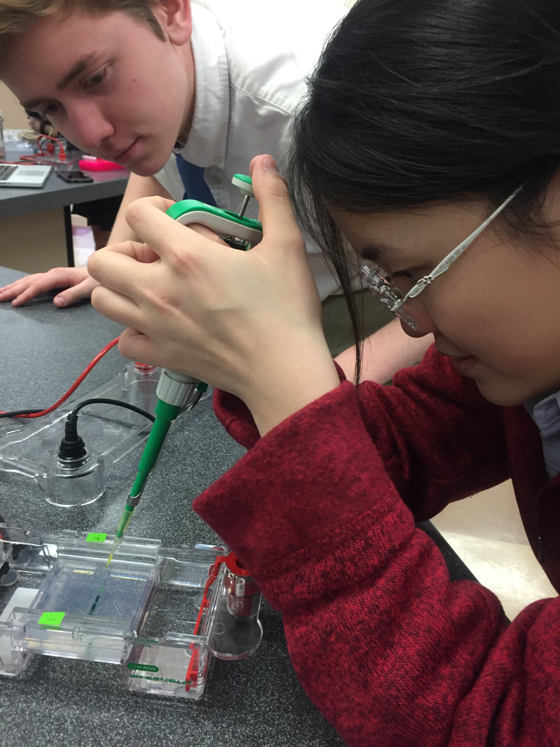
Lab#1: Measurements, Micropipetting, and introductory Electrophoresis for DNA Labs Lab partners: Vaishnavi Kumar, Gray Martucci. Introduction: The purpose of this lab is to learn several important techniques performed in DNA labs such as using micropipettor to transport small amount of liquid, casting the gel, loading the wells accurately using micropipettor, and gel electrophoresis. Although we used candy dye instead of actual DNA in this lab, the basic concept of how the dye elements are separated by colors is the same as how DNA segments are separated by different sizes. Similarly, both the dye and the DNA samples are loaded into the wells, and “an electric current is applied to pull them through the gel. DNA fragments are negatively charged, so they move to the positive electrode. Because DNA fragments have the same amount of charge per mass, smaller fragments move through the gel faster than larger ones.”(Gel electrophoresis) This technique helps scientist visualize DNA fragments since they can see the groups of DNA fragments with the same size in bands. “Using electrophoresis, scientists can understand how many different DNA fragments are present in a sample and how large they are relative to one another.”(Gel electrophoresis) Source cited: "Khan Academy." Khan Academy, 2018, https://www.khanacademy.org/science/biology/biotech-dna-technology/dna-sequencing-pcr-electrophoresis/a/gel-electrophoresis. "What Is Gel Electrophoresis?." Yourgenome.Org, 2018, https://www.yourgenome.org/facts/what-is-gel-electrophoresis. Lab procedure: 01.16.18 B Tue. Preparing the gel, extracting the food dye from candies, and preparing four samples with standard color dye: We start with pouring the melted agarose gel into the gel tray wrapped with electrical tape, put the comb in it to cast 9 wells. When are waiting for the gel to solidify, we use dye extraction solution to get the food dye on the M&M candy coating. Once we get the dark solution, remove the candy from the cup. Pipette the solution into labeled microcentrifuge tube. Use a micropipettor size 2-20µL, we prepare four sample tubes A, B, C, and D each with different amount of four standard dyes, Blue 1, Yellow 5, Yellow 6(orange), and Red 40. Centrifuge all the test tubes and sit them on a tube rack. 01.17.18 C Wed. Loading the gels and Gel electrophoresis: First we take out the comb from the gel, put the solidified agarose gel with the tray into a gel rig, pour a 0.892 TAE buffer so all parts of the gel are immersed under the buffer. Use micropipettor size 2-20µL, load around 10-15µL of each sample carefully into each wells from left to the right(label different sides of the gel rig). Close the lid of the gel rig, plug it into power source and start electrophoresis. Starting time: 10:28am, with a voltage of 100V. After around 17 min, we observe that most of our samples have traveled obvious distance to the positive side, we turn off the power source and end the electrophoresis. We take the gel out of the gel tray and take a picture of the gel on the light screen. Results: Data table: (we print out the photograph and measure from paper.)   Conclusion:
In the four sample test tube, A, B, C, and D, blue color dye bands all appear which matches our prediction. They are also the closest to the wells, which shows that blue dye molecules have the biggest size among all. Below all the blue bands, well A, B, and D show orange bands, too, which matches our table since we add yellow 6(orange) dye to test tube A, B and D. Close to the orange dye, only well C shows an obvious red band while we did add red 40 dye to test tube A, C, and D. An educated guess will be in test tube A and D, yellow 6(orange) may travel to a very similar distance to the red 40 and our eyes can't pick up the color difference. All the dye molecules in our seven samples migrate towards the positive electrode, meaning the molecules are negative. The dye molecules which travel the fastest are the yellow dye molecules; the dye molecule travel the shortest are the blue molecules. The distance traveled varies by different type of the molecules. This is because shorter, smaller molecules travel through the agarose gel pores faster that long, bigger molecules do. In both my lab partner Gray and Vaishnavi’s candy samples, yellow 5 dye bands appear. The results matches our M&M candy colors we chose, too. Gray picks a green one, and blue and yellow bands show up; Vaishnavi picks a yellow candy, and only yellow band appears; I chose a orange candy, so only yellow 6(orange) dye shows up. Discusion: The electric current that runs through the buffer and the gel forces the charged molecules in the gel to move to opposite charged ends. In DNA molecules, the charge and the size influence how the dye molecules migrate in an agarose gel. Also, the agarose gels have the right size of dense pores that allows molecules to squeeze through if they are small enough, while bigger molecules are trapped and stay at the same place and form a “band” which can be visible to us. The charge changes the direction of the migration. Negatively charged dye molecules will be pulled towards the positive electrode, while some positively charged molecules will travel to the negative side. The size influence the distance traveled by the molecules, the smaller the molecules are, the faster they travel through the agarose gel pores, resulting in longer distance traveled, and vice versa. Among the carminic acid, betanin, fast green FCF, and Citrus Red 2 those four dyes, the Fast green FCF will be the most suitable for our lab. Since this dye has a negative charge, it will move towards the positive end of the gel and makes it more obvious for us to see. To prepare for our color dye gel electrophoresis experiment, we cast the wells in the middle of the gel. Different from color dyes molecules that may be negatively or positively charged, the DNA molecules are all negatively charged. When we are using DNA molecules to perform the gel electrophoresis, we only have to cast the wells at one end, and face it towards the positively charged electrodes in order for all DNA molecules to run towards the positive end. Dalton is a unit for atomic mass. DNA molecules with larger daltons like 5000da will move slower to the positive end of the gel, resulting in a short distance from the well. DNA molecules with a smaller daltons like 600da will move faster, resulting in a long distance far away from the wells. The electrophoresis allow the scientists to directly measure the daltons of DNA fragments by calculating their distance traveled, therefore, scientists can compare different sizes of DNA fragments. Bibliography: Works cited: "Khan Academy." Khan Academy, 2018, https://www.khanacademy.org/science/biology/biotech-dna-technology/dna-sequencing-pcr-electrophoresis/a/gel-electrophoresis. "What Is Gel Electrophoresis?." Yourgenome.Org, 2018, https://www.yourgenome.org/facts/what-is-gel-electrophoresis. "Unified Atomic Mass Unit." En.Wikipedia.Org, 2018, https://en.wikipedia.org/wiki/Unified_atomic_mass_unit.
3 Comments
Jack Grossman
2/2/2018 08:02:27 am
This lab report was really well done, the detail and research elements were especially impressive. One thing you could add is a discussion on the errors and potential improvement of the lab at hand.
Vaishnavi
2/2/2018 08:04:37 am
Elaine!! I love how you included the dates in the methods section cause it makes more personal. It's more interesting to read a daily log than a long paragraph.
Olivia S
2/2/2018 08:13:46 am
Great job Elaine! I loved how thorough you were with all of your information. Your pictures were so fun! You were precise and made sure to walk the reader through the lab. Your blog is so cute! I love it! Your comment will be posted after it is approved.
Leave a Reply. |
AuthorHello, I am Elaine. A junior at Holland Hall School. I am passionate about learning genomics and genome editing. In this blog page, you will see lab reports, personal opinion pieces, and some essays on biology! Archives
April 2018
Categories
|


 RSS Feed
